What is Peripheral Vascular Disease?
Peripheral Vascular Disease (PVD) also referred to as peripheral artery disease is a common disease that occurs when the blood vessels that supply blood to the limbs and other organs of our body are partially or completely blocked due to plaque build-up, a condition called atherosclerosis. Plaque forms out of the substances present in the blood such as fat, cholesterol, calcium and fibrous tissue. These plaque deposits gradually harden and narrow down the arteries. This limits the oxygen-rich blood supply to the various parts of your body. The most commonly affected blood vessels because of PVD are the arteries of legs.

Symptoms Of Peripheral Vascular Disease
- Difficulty in walking
- Pain and muscle cramps in the legs while walking or climbing stairs (intermittent claudication)
- Numbness, weakness or heaviness of muscles
- Cold sensation of the skin in legs or feet
- Discoloration of the skin, predominantly in arms or legs
- Sores on toes and feet, which are difficult to heal
- Burning or aching sensation in feet and toes
Test for Peripheral Vascular Disease
Peripheral vascular disease screening helps in detecting early signs of PVD that can be potentially treated before further deterioration of the condition. Peripheral vascular disease screening involves painless and non-invasive tests, which include:
- A medical and family history
- Medication review
- Leg pain assessment
- Blood pressure reading
- Body fat analysis
- Body mass index
- Cholesterol panel
- Blood glucose estimation
- Lipid profile
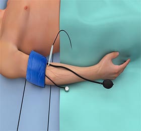
- Ankle-brachial index -Ankle-brachial index (ABI) compares the blood pressure of your ankle to blood pressure in the arm.
- Doppler ultrasound- This is done to check for a blocked artery, where blood flow through the artery will be determined. It also helps in determining the severity of PVD.
- Treadmill Test - This test is done to gauge the severity of symptoms and to monitor the level of exercise that elicits the symptoms.
- Magnetic Resonance Angiogram – Magnetic resonance angiogram (MRA) makes use of magnetic and radio waves to produce images of blood vessels. It can detect the location of a blockage in the blood vessel.
- Arteriogram - This test locates the blocked artery. The test involves the injection of a dye into your artery, followed by which an X-ray is taken. X-ray shows the location and extent of blockage in the artery.
- Diabetes Mellitus
- High blood pressure
- Obesity
- Lack of exercise
- Smoking
- High blood cholesterol
- Family history of atherosclerosis and circulatory problems


